Sunday, 12 July 2020

Quantitative Fluorescence PCR (QF-PCR) in prenatal aneuploidy testing

Quantitative Fluorescent PCR is a PCR-based technique in which a low number of PCR cycles are used, such that the endpoint of the reaction reflects the DNA or chromosome copy number in the test sample.
There are two key steps to QF-PCR, amplification and quantification. Firstly, specific regions of DNA are amplified. And then secondly, the amount of DNA present in those regions is quantified.

We will first concentrate on the initial QF-PCR step, the amplification of DNA. QF-PCR exploits DNA regions containing specific DNA markers or Short Tandem Repeats, or STRs for short, that are scattered throughout the genome.
These STRs are repeat sequences of tetranucleotides, or four bases, and are highly polymorphic in terms of their number of repeats, so that and individual is highly likely to have a different number of repeats on each homologue. Primers are designed for each marker. These primers are labelled with a fluorescent tag. The repeats are then amplified by PCR, allowing comparison of the relative dosages of maternal and paternal chromosome and against those of a normal control. Only those STRs or markers that differ from each other are chosen for analysis, so-called informative markers. The example here shows an STR which has six repeats on the maternal chromosome 21 and four repeats on the paternal chromosome 21, and is therefore informative.


The marker regions are amplified in a PCR reaction. However, there is a very important difference in the PCR reaction used for QF-PCR. That is, that the reaction is stopped early in the exponential phase, generally between 24 and 26 cycles. This is because the assumption is that within the early exponential of amplification, the amount of product present is directly proportional to the amount of target sequence present in the original template. Therefore, the results indicate the quantity of original template present. The success of the assay is therefore dependent on the amount of DNA used in relation to the number of amplification cycles. 

The second part of the QF-PCR experiment involves quantification of the DNA.
In this phase, the PCR products are separated by capillary electrophoresis. Lasers excite the fluorochromes, and the relative amount of fluorescence emitted, which is proportional to the amount of each amplified allele present, is measured.

QF-PCR is used to confirm copy number variants detected by array CGH, MLPA, or Next-Generation Sequencing read depth analysis.
However, one of its primary uses, given the speed of results and associated low cost, is in prenatal aneuploidy testing. We will focus on this use of QF-PCR for the remainder of this presentation. 
Prenatal aneuploidy testing can be undertaken on CVS or amniotic fluid samples. It has a fast turnaround time, and the result is available within three to five days.

Analysis is done on markers from chromosomes 13, 18, and 21, and may also be done on markers from the sex chromosomes. This diagram shows a series of markers across chromosomes 13, 18, and 21 that are used for aneuploidy testing. The blue markers are the primary markers, which are analysed first. And the red markers are additional markers, which can be used for confirmation of a result, or if the primary markers are uninformative. A pair of primers is designed for each STR to be run, and multiple pairs of primers can be run in a single reaction. In this way, multiple loci are screened across multiple chromosomes simultaneously.

Analysis is done on markers from chromosomes 13, 18, and 21, and may also be done on markers from the sex chromosomes.


This diagram shows a series of markers across chromosomes 13, 18, and 21 that are used for aneuploidy testing. The blue markers are the primary markers, which are analysed first. And the red markers are additional markers, which can be used for confirmation of a result, or if the primary markers are uninformative. A pair of primers is designed for each STR to be run, and multiple pairs of primers can be run in a single reaction. In this way, multiple loci are screened across multiple chromosomes simultaneously.
Markers can also be used across the X and Y chromosomes to check the sex chromosome complement. Analysis software is used to compare peak heights between different alleles of each STR.
The left-hand diagram shows a normal diallelic sample, that is representing markers from two chromosomes. The top marker is homozygous in this sample, giving only one big peak, which is uninformative. The marker below is heterozygous, and in this normal sample gives a 1:1 ratio. The right-hand diagram shows a triallelic sample, resulting from trisomy. Again, the top marker is uninformative, as all three alleles have the same length STR. 
The middle marker shows two peaks in an abnormal 2:1 ratio, reflecting the presence of two copies of one of the parental alleles. The bottom marker shows an abnormal triallelic result of 1:1:1. This reflects the presence of three different parental alleles, likely to have resulted from meiotic nondisjunction during oogenesis. 
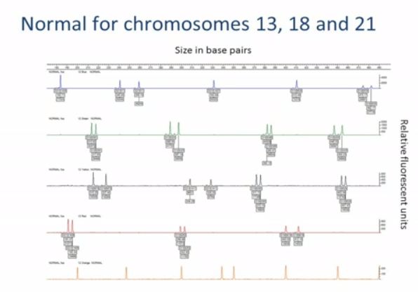
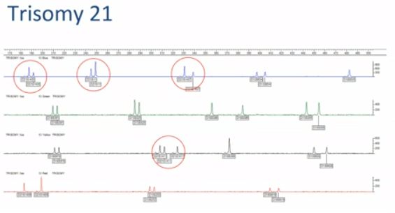
Results from multiple markers are represented graphically for analysis. The ratios are then also displayed in an Excel spreadsheet. This graph shows the result from a sample that is normal for chromosomes 13, 18, and 21. In the blue trace, you can see some uninformative homozygous markers, giving just a single peak. In the green, black, and red traces, you can see that the marker peaks are paired,and their heights are in a normal 1:1 ratio. This graph shows a sample with trisomy 21.

Here, you can see that the chromosome 21 markers, ringed, are present in abnormal 2:1 or 1:1:1 ratios. The other markers, for chromosomes 18 and 13, are present in normal 1:1 ratios. 
This result shows mosaicism for trisomy 18. The boxed peaks are the chromosome 18 markers. Most of them appear normal, but if you look carefully at the boxed peaks on the top blue trace, you can see a small third peak in the middle. This subtle change indicates the presence of a third allele at this chromosome 18 marker. The level of mosaicism in this sample is around 15%, which is usually the minimum limit of detection. As with all prenatal samples, especially CVS samples, confined placental mosaicism can be a problem. A triallelic result indicates the meiotic nondisjunction event during oogenesis, so carries a high risk of foetal involvement.
However, a diallelic result when none of the markers confirm nondisjunction raises the possibility that the cell line may be confined to the placenta. In all cases, the QF-PCR result should be carefully interpreted in conjunction with the ultrasound findings. Any abnormal results on CVS are further investigated with a karyotype, and further testing with amniocentesis is often undertaken if the CVS results are ambiguous. 

Maternal cell contamination is a common problem with prenatal samples. It is detectable by skewed ratios or extra peaks involving all the chromosomes, as shown here. This sample was reported as unsuitable on the basis of the evident maternal cell contamination. In summary, QF-PCR involves the amplification and quantification of markers on chromosomes of interest. 
QF-PCR is used to confirm copy number variants detected by other methods. However, its main application is in prenatal aneuploidy testing as it is a quick, reliable, and cost-effective test.
Source:-





Saturday, 11 July 2020

Non-invasive prenatal diagnosis (NIPD)

Non-invasive prenatal diagnosis (NIPD) uses the mother’s blood rather than the fetal genetic material when making prenatal diagnosis of genetic conditions.


NIPD for sex determination

NIPD for fetal sex determination can be undertaken from 7 weeks gestation for pregnancies at a high genetic risk.
Common referral indications are pregnancies at risk of serious X-linked conditions or congenital adrenal hyperplasia, or where there are ultrasound findings such as ambiguous genitalia.
Some common examples of the application of this technique in the clinic include:
  • Fetal sexing in pregnant women who are carriers for an X-linked disorder such as Duchenne Muscular Dystrophy (DMD) or haemophilia. In such cases, early fetal sex determination can reduce the invasive diagnostic testing rate by around 50% (as female fetuses may not require further testing).
  • Fetal sexing for couples where both parents are carriers of congenital adrenal hyperplasia (CAH). Early identification of a male fetus can avoid unnecessary fetal exposure to steroids as only female fetuses require steroid treatment to avoid virilisation.
For fetal sex determination, ultrasound scanning must first be undertaken to confirm the gestation, and to identify multiple pregnancies or empty sacs from early twin demise, which could confound the results.
Once the cfDNA has been extracted, it is analysed using real time quantitative PCR methods to look for Y chromosome markers, indicating a male pregnancy.
Absence of Y signal indicates a female pregnancy.

NIPD for monogenic disorders

NIPD can be used for some monogenic disorders to exclude either paternal or de novo alleles (i.e. alleles that are not present in the mother). The techniques can be used in both dominant conditions, or recessive conditions where the parents are carriers of different mutations.
NIPD has been worked up for certain monogenic disorders such as skeletal dysplasias, congenital adrenal hyperplasia and cystic fibrosis, but bespoke tests can also be designed for other single gene disorders, especially in the context of testing for a subsequent pregnancy for a mutation previously identified by invasive prenatal or postnatal testing.
A) NIPD for dominant conditions – the exemplar of skeletal dysplasia
NIPD has been approved for prenatal diagnosis of FGFR3-related skeletal dysplasia (achondroplasia and thanatophoric dysplasia) in the NHS since 2012.
Achondroplasia is the most common short stature syndrome, presenting in the 3rd trimester. It shows autosomal dominant inheritance, although often arises de Novo.
A single mutation is responsible for 98% of cases. cfDNA testing can, therefore, be undertaken using a mutation-specific PCR-based restriction enzyme digest. NIPD allows confirmation of a diagnosis made on ultrasound, excluding other more severe dysplasias without the risk of preterm labour.
Thanatophoric dysplasia (TD) is the most common lethal skeletal dysplasia. It arises de novo and may be caused by at least 13 different mutations in FGFR3.
NIPD enables early diagnosis, allowing the option of surgical termination if requested (which would not be possible later in the pregnancy), and allowing accurate diagnosis in twin pregnancies without putting an unaffected twin at risk.
Initially, NIPD for TD was also developed using restriction enzyme digest PCR to identify the most common mutations, however since it can be caused by such a variety of possible mutations, less costly and time-consuming testing using a next-generation sequencing gene panel was developed, which is now being used for all FGFR3-related skeletal dysplasias.
NIPD can also be used in subsequent pregnancies following a pregnancy affected by an apparently de novo mutation, to exclude the possibility of recurrence resulting from germline mosaicism.
B) NIPD for recessive conditions – the exemplar of cystic fibrosis
NIPD can be used in recessive conditions where couples are known to be carriers of different mutations.
In this case, testing is done to exclude the presence of the paternal mutation, which means that the fetus is either a carrier (of only the maternal mutation), or completely unaffected.
If a fetus is shown to have the paternal allele, then they might be either a carrier of only the paternal mutation, or affected, and invasive testing will be required to distinguish this. Nevertheless, paternal mutation exclusion allows for a reduction in invasive testing.
In the case of cystic fibrosis, NIPD can currently be used where the couple are each known to be carriers of different mutations, and the paternal mutation is one of the 10 most common mutations covered by the NGS panel used for analysis.
NIPD for cystic fibrosis has been approved under these circumstances in the NHS since 2014.

Future directions

Current research in NIPD is now focusing on how to extend its use in order to detect a mutation in the fetal DNA against the background of the same mutation in the mother.
These approaches depend on precise measurement of relative mutation dosage (RMD). If the mother is heterozygous for the location of interest, then the presence or absence of the same mutation in the fetus will affect the expected extent of allelic imbalance between mutant and wild-type DNA in the total cfDNA (i.e. maternal and fetal cell-free DNA combined).
But the concentration of cfDNA varies between samples so this approach requires a precise quantification of the cfDNA by measuring a fetal allele not present in the maternal genome. This approach is extremely technically demanding and at present is only in the research phase.
Source:- 

Saturday, 27 June 2020

NIPT Or Non-invasive prenatal testing for aneuploidy

Non-invasive prenatal testing (NIPT) for aneuploidy is rapidly becoming the first line screening test for trisomies 13, 18 and 21 in the first trimester, with more and more hospitals offering the test as part of their routine prenatal care.
Three main methods are used in NIPT. Each is associated with specific advantages and disadvantages and we will discuss each in turn.
  • Massively Parallel Shotgun Sequencing (MPSS)

This approach was described by Fan et al. in 2008 (PNAS, 1056(16):266-71) and uses next-generation sequencing technologies to generate reads representing chromosome regions across the genome.
If there is trisomy of a particular chromosome, this would be detectable by a relatively greater number of reads mapping back to that chromosome region on the reference genome. However, this relative increase might be very small indeed.
For instance (as reported by the NIPT diagnostic laboratory at the South West Thames Regional Genetics Service) the reads representing chromosome 21 in a normal pregnancy typically might constitute 1.36% of the total cfDNA (remember, the total cfDNA is maternal and fetal cell free DNA).
However, in a pregnancy affected with trisomy 21, the total contribution of reads mapping to chromosome 21 from both mother and fetus might increase to just 1.42%. That is only a 0.06% difference!
Therefore, to distinguish such small differences in the amount chromosomal DNA found, an incredibly accurate counting and sorting method is required.
A disadvantage of MPSS is that, depending upon the underpinning NGS technology, the method can be costly with only a relatively small proportion of the generated reads being analysed.
  • Targeted multiple parallel sequencing (MPS)

There are various different forms of targeted MPS. However, they are all underpinned by the same concept - amplifying and quantifying chromosome-specific sequences.
One form of targeted MPS is DANSR (Digital Analysis of Selected Regions). DANSR uses oligonucleotide probes that hybridise to target sequences.
The principle is a bit like that behind MLPA in that it is the probes that are amplified rather than the target sequence. However, in contrast to MLPA, DANSR used three rather than two probes.
The number of each of the amplified probes reflects the amount of target sequence. Therefore, where a trisomy is present, there will be a relatively greater number of amplified oligonucleotides.
An advantage of targeted MPS is that costs are reduced because fewer sequences are generated. However, the targeted sequencing introduces biases and some believe reduces accuracy.
  • SNP detection

SNP detection is very different from the above two methods as, rather than analysing cfDNA as a whole, it differentiates between maternal and fetal cfDNA. In the commercially available kits, about 20,000 SNPs are utilised.
First of all, the maternal SNP genotype is determined through analysis of the buffy coat (the buffy coat is a layer of blood, mostly containing leukocytes and platelets and therefore not containing cfDNA, obtained through the centrifugation of a whole blood sample).
The plasma is then SNP genotyped which represents both fetal and maternal SNPs. Computationally, the two SNP results are compared and individual fetal and maternal SNP profiles are calculated.
Fetal fraction is first calculated. For instance, if mum is AA and fetus is AB for a given SNP locus, if there is 1000 of AA to 100 AB, then the fetal fraction is 10%.
Then, in order to determine copy number for each interrogated chromosome locus, the maternal and fetal genotypes are compared and evaluated to see whether there is more or less than expected.

Accuracy of NIPT

NIPT has been shown to have high sensitivity and specificity (>99%) for trisomy 21 detection. These figures are currently slightly lower for the other trisomies and for sex chromosome imbalance detection.
It is important to remember that these tests should be considered an advanced screening test rather than a diagnostic test. Sensitivity (likelihood of detecting a trisomy if present) is not the same as positive predictive value (PPV; likelihood of a positive result being a true positive), and the PPV for these tests are rather lower than the sensitivities.
Thus any positive result must be confirmed by invasive testing before any management decisions are made for the pregnancy.
The negative predictive value (NPV; likelihood of a negative result being a true negative) is high for these tests, so in the context of normal ultrasound findings a negative test is reassuring, and confirmation with an invasive test is not usually done.
However, in the context of abnormal findings on ultrasound, a negative NIPT should not be considered reassuring as other underlying chromosomal abnormalities have not been excluded and further invasive testing should be considered.

clinical situations is cell-free fetal DNA analysis not possible for detection of a maternally inherited single gene mutation.
NIPD cannot distinguish the presence or absence of a maternally inherited mutation in the fetus. This is because the mother carries the same mutation, so it is present in both the fetal and maternal cell free DNA fractions in the maternal plasma. Researchers are working on developing techniques which analyse the dosage of a given mutation in the maternal blood, but this is technically highly demanding and not currently developed for clinical use.

Twin pregnancy is not likely to cause an inconclusive result when performing NIPT for aneuploidy.
NIPT can be performed in twin pregnancies and is usually informative. If the aneuploidy screening comes back negative then this applies to both fetuses, and in the absence of any other indications, invasive testing can be avoided. If NIPT shows a possible aneuploidy then it could be difficult to know whether one of both foetuses is affected, and invasive testing is indicated.

Cell free DNA (cfDNA)

Cell free DNA (cfDNA) analysis is revolutionising prenatal genetic testing. Rather than subjecting women to invasive tests with associated discomfort and increased risk of miscarriage, analysis of cfDNA (through non-invasive prenatal testing, NIPT and non-invasive prenatal diagnosis, NIPD) is offering pregnant women an earlier, safer alternative.

Because of the close contact between maternal and fetal circulations in utero, a small amount of cell-free fetal DNA (not encased in a cell or nucleus) enters the maternal blood stream.
It originates from the trophoblast (placenta). This can be detected and analysed by taking a blood sample from the mother. Fetal DNA makes up around 10-20% of the total circulating cell free DNA in the maternal plasma. The cell free DNA in the maternal plasma comprises both maternal and fetal cell free DNA fractions, and distinguishing one from the other is one of the main challenges of cell free DNA analysis.
The presence of cell-free fetal DNA in maternal plasma and serum was first described by Lo and colleagues in the Lancet back in 1997.
Lo also spoke about how the arrival of whole genome sequencing brought about groundbreaking developments in the field of prenatal diagnosis at the PHG Foundation 15th anniversary conference, ‘Translating genomics: making science work for health’ in 2012.
The earliest stage in gestation that cell-free fetal DNA can be detected 4-5 weeks gestation. How soon after delivery do the cell-free fetal DNA levels fall. It is cleared from the maternal circulation within 30 minutes of delivery.
NIPD refers to the use of cfDNA analysis to diagnose monogenic disorders or to determine fetal sex or rhesus status. It does not require an invasive test for confirmation of the result.
NIPT refers to the use of cfDNA analysis as an advanced screening test for aneuploidy. Whilst this provides an accurate screening test for aneuploidy from 10 weeks’ gestation, positive results require confirmation by invasive testing.

Karyotype methods and applications


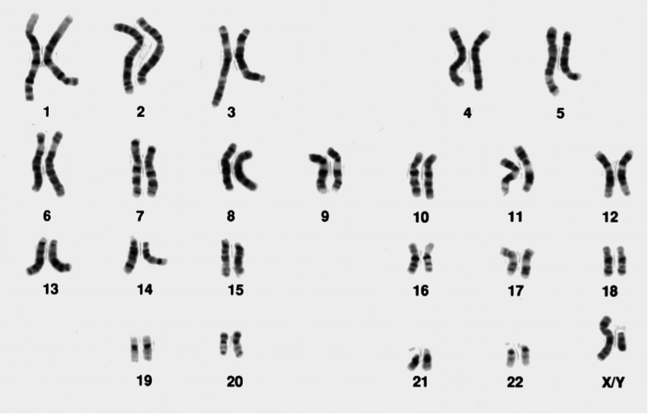
A karyotype describes all the chromosomes of a cell, usually visualised as a systemised arrangement of chromosome pairs, in descending order of size.
It is used to analyze the number and structure of all the chromosomes and provides a low-resolution genome-wide screen for chromosomal abnormalities.
An image of a normal male karyotype with 23 pairs of chromosomes
Figure 1: A normal male karyotype (note the X and Y sex chromosomes)
Original image from the National Institutes of Health and used under a public domain license.
When clinical cytogenetic analysis started in the 1960s, solid staining was used, which meant that the different chromosomes were only recognizable by size and shape.
In the 1970s the visualisation of chromosomes improved due to the development of specific stains and enzyme digestion techniques, giving the chromosomes reproducible banding patterns. Different techniques were utilised by different countries.
For instance, France used Q-banding whereas the UK used a combination of enzyme digestion with trypsin and G-banding (see below). Using these techniques, each chromosome pair could be visualised with a unique pattern, resembling a bar code.
This not only helped cytogeneticists to recognise the different chromosomes, but also allowed them to see when parts of the chromosome were rearranged, deleted or duplicated. The most widely used stain is the Giemsa stain, giving the familiar G-banding appearance shown in Figure 1.

How is a karyotype done?

Karyotyping can be done from any sample from which you can obtain nucleated cells which retain the potential to divide, most commonly:
  • Blood
  • Bone marrow
  • Skin
  • Prenatal samples (such as CVS or amniotic fluid)
Cells are cultured in an environment which includes stimulants for cell division, then arrested at metaphase and harvested for analysis. Cell culture can take anything from 3 days (blood/bone marrow) to 7-14 days (skin, prenatal samples).
This requirement to culture the cells increases the turnaround time, but is unavoidable.
Once harvested, the cells are mounted on slides, banded (enzyme treated and stained), then viewed under a light microscope (x1000).

How is a karyotype analysed?

The cytogeneticist counts the chromosomes, pairs the homologues and compares the banding pattern in each set of homologues to identify any differences. They are aided by software which allows them to ‘cut and paste’ the chromosomes so that they can be aligned for comparison, as shown in Figure 1.
Many attempts have been made to develop computer software to replace the job of the trained cytogeneticist but it has not been possible to replace the pattern-recognition skills of the human eye.
Even in the best hands however, the limit of resolution of a karyotype is around 5 Mb, and in many applications karyotyping has been superseded by the use of higher-resolution techniques such as array-CGH.

What sorts of abnormalities can a karyotype identify?

Karyotyping is able to identify numerical abnormalities in the chromosomes (also known as aneuploidy), where a chromosome is missing or present in one or more extra copies. Examples of this include trisomy of chromosomes 13, 18 or 21, or sex chromosome abnormalities such as Turner syndrome (45,X) or Klinefelter syndrome (47,XXY).
Karyotyping can also identify structural abnormalities in the chromosomes including rearrangements (such as translocations, insertions or inversions), deletions and duplications.
Chromosome deletions can be detected on karyotype down to a resolution of around 5 Mb, but some are very subtle and require a trained eye.
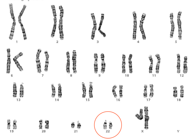
The karyotype shown in Figure 2 shows a 22q11.2 deletion associated with velocardiofacial (or DiGeorge) syndrome. Would you have spotted it?
Image of a Karyotype showing 22q11.2 deletion
Figure 2: Karyotype showing 22q11.2 deletion
© St George’s, University of London

How are karyotypes reported?

A clinical karyotype report will include a description of the cytogenetic findings, described using standardized nomenclature and displayed using a cartoon representation of the chromosome and its banding pattern, called an ideogram (Figure 3).
Image of an ideogram representation of chromosome 13
Figure 3: Ideogram representation of chromosome 13 Original image from Human chromosome ideograms from NCBI’s Genome Decoration Page 
There may also be a description of any additional tests undertaken to confirm the results, a literature review with any suspected associations with phenotype, a description of any follow-up studies required and, where appropriate, recurrence risks will be given.

What is karyotyping used for currently?

In many contexts karyotyping has been superseded by the use of array CGH, which can also pick up numerical and structural abnormalities in the chromosomes, and at much higher resolution, however karyotyping is still in use in certain clinical situations.
One such situation is in the investigation of infertility or recurrent miscarriage, where a balanced rearrangement is suspected. A balanced translocation describes a rearrangement of chromosomal material, but with no overall gain or loss of genetic material.
The balanced translocation carrier is usually unaffected themselves, however problems may arise when they try to have children as the balanced translocation can affect the way that the chromosomes divide during meiosis, and imbalance can then occur in the offspring.
In this context, karyotyping has a distinct advantage over array CGH in that it is able to detect balanced rearrangements, which array CGH is not.
This is because a karyotype provides a visual representation of the chromosomes, unlike array CGH, which provides quantitative information on the amount of chromosomal material present, but not its location.

Question 1

1. Which of the following samples is not suitable for karyotype analysis?
2. Which of the following statements about karyotype analysis is false?
3. Study the karyotype below. Can you spot the diagnosis?
4. Study the image below which includes a chromosome deletion associated with a recognised developmental disorder. Compare each of the chromosomes to its pair to see whether you can recognise any difference in banding pattern which might be indicative of there being a chromosome deletion or duplication. What syndrome do you think this patient has?

Answers:-
1. D
An EDTA tube cannot be used. The correct blood tube to use for karyotype analysis is lithium-heparin. Samples for array CGH are collected in an EDTA tube however.

2. B
For karyotype preparation chromosomes are arrested in metaphase.

3. D
One copy of chromosome 22 looks shorter, and one copy of chromosome 9 appears longer. This translocation gives rise to an oncogenic fusion gene bcr-abl, causing chronic myeloid leukaemia (CML).

4. D
The short arm of the left-hand chromosome 4 is shorter in the top image than in the bottom image, due to a 4p deletion. This is associated with Wolf Hirschhorn, a developmental disorder characterised by a distinctive facial appearance, a (usually) severe intellectual disability and sometimes seizures.
Source:-


Rare Diseaes Some examples of rare diseases are  Spinal Muscular Atrophy ,   Osteogenesis imperfecta ,   Achondroplasia   or   Rett Syndrome...